Splicing’s final act: Three molecular hands untying a knot
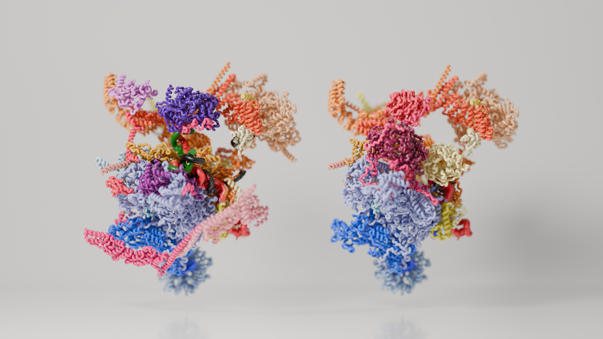
The correct expression of genes is essential to produce the vast diversity of proteins needed for life. A crucial moment in gene expression is splicing, during which a large molecular machine—the spliceosome—edits RNA before it is used for protein production. For each editing task, this intricate machine must assemble, complete its work, and rapidly come apart; but exactly how it reaches the end of this cycle has been a missing piece of the puzzle.Researchers led by Clemens Plaschka at the IMP have now uncovered the spliceosome’s hidden final act, revealing how three molecular motors work together to dismantle the machine and complete its termination—redrawing the textbook pathway of this splicing stage. The study is published in the journal Nature.
The life of a cell is sustained by a constant flow of information from genes to proteins. This process, known as gene expression, underlies the production of a plethora of different proteins which form cells that grow, adapt, and carry out specialised roles. To produce a protein, cells first copy the relevant genetic information from DNA into precursor messenger RNA, or pre-mRNA—a preliminary blueprint for protein production. Before it can be used, however, this blueprint must be carefully edited.
For this task, cells rely on the spliceosome— an extraordinarily complex molecular machine built from five RNAs and more than 100 proteins. The spliceosome edits pre-mRNA by cutting out non-coding regions called introns and stitching the remaining coding regions, or exons, together. For every intron, the spliceosome assembles from scratch and repeatedly reshapes itself as it cuts and reconnects the RNA, ultimately producing mature messenger RNA (mRNA)—a continuous, readable set of instructions for protein production.
Once the mature mRNA has been produced, the spliceosome must reset for the next editing task. But first, it has to discard the discarded intron still tangled within its core and then come apart, allowing its components to be used again. These final steps unfold so rapidly that scientists have been unable to catch the spliceosome in the act and understand the events at the molecular level.
Researchers led by Clemens Plaschka at the IMP have now revealed what happens as the spliceosome prepares to be recycled. By isolating spliceosomes directly from human cells and examining them with cryo-electron microscopy, the team have captured these fleeting stages that scientists had been unable to observe before. They discovered two previously unknown states of the spliceosome and showed how three molecular motors progressively untangle the machine from within. The findings, published now in the journal Nature, reveal a missing chapter in one of biology’s fundamental processes and redraw the textbook pathway of splicing.
A hidden final act
Taking apart the spliceosome is a delicate operation. After editing the pre-mRNA, the machine must separate the discarded intron from a dense network of RNA and proteins, dispose of it and recover its own components for reuse. Because this process unfolds rapidly and its intermediate stages are fleeting, scientists had been unable to follow the spliceosome through this crucial part of its life cycle.
To tackle this challenge, the Plaschka lab drew on a particular strength: their lab’s experience in isolating intact molecular machines as they naturally form inside human cells. The researchers grew large volumes of human cells to recover enough of the rare, short-lived spliceosomes that accumulate only briefly as the machine comes apart. This approach allowed them to search for intermediate states without having to predict and recreate them in the laboratory. Using cryo-electron microscopy, they then determined their three-dimensional structures, including the highest-resolution structure of an assembled human spliceosome to date.
“We study native molecular assemblies because it allows us to investigate molecular events that may escape more classical reconstitution approaches,” says Clemens Plaschka. “In this case, we captured states of the spliceosome that we did not know existed.”
Together with the lab of Stefan Ameres at the Max Perutz Labs, the researchers used a specialised RNA-sequencing approach to confirm the identity of the newly discovered state. It captured the spliceosome midway through its final act: the discarded intron had been opened, but had not yet been released.
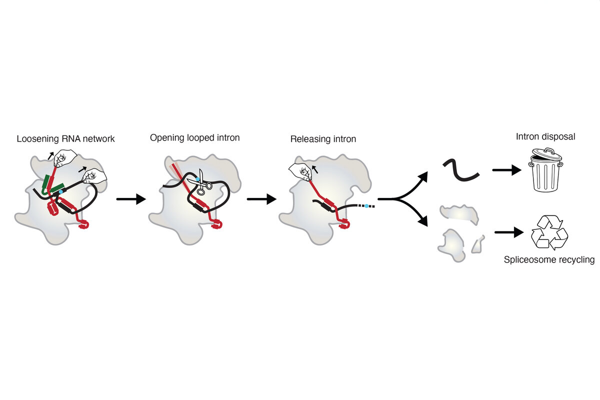
What happens to the spliceosome resembles untying a tightly knotted pair of shoelaces. The researchers found that three RNA helicases—molecular motors that pull and rearrange RNA—act in sequence to loosen the spliceosome from within.
“We thought that a single helicase would take the spliceosome apart,” says first author Vytaute Boreikaite. “Instead, we found three—like three hands pulling on different parts of a knot. Together, they loosen the RNA network, help to open the discarded intron and finally free it from the spliceosome.”
This leads to a new model for how splicing ends. Two of the molecular motors first loosen the knot, allowing the looped intron to be opened and prepared for disposal. The third then pulls the intron free, bringing the spliceosome to the point of collapse. Rather than occurring separately, the disposal of the intron and the dismantling of the spliceosome are
therefore closely linked.
The findings also offer clues to how cells dismantle spliceosomes that become trapped before completing their task. Regular and faulty spliceosomes appear to rely on much of the same machinery, providing researchers with a new foundation for investigating how cells safeguard splicing. By uncovering this previously hidden part of the process, the study fills in the textbook pathway of gene expression.
“Splicing is fundamental to the enormous diversity of proteins that cells and organisms can produce,” says Plaschka. “Understanding this machinery ultimately brings us closer to understanding how messenger RNA is made and how biological complexity is built .”
Original Publication
Vytaute Boreikaite, Rupert Faraway, Matthias K. Vorländer, Alexander W. Phillips, Leonie Opitz, Moritz Wanke, George Yakoub, Gerald Raffl, Laura Fin, Román González-Prieto, Martijn S. Luijsterburg, Stefan L. Ameres, and Clemens Plaschka. “Mechanism of spliceosome termination.” Nature. DOI: 10.1038/s41586-026-11101-6
Further reading
About the IMP at the Vienna BioCenter
The Research Institute of Molecular Pathology (IMP) in Vienna is a basic life science research institute largely sponsored by Boehringer Ingelheim. With over 220 scientists from 40 countries, the IMP is committed to scientific discovery of fundamental molecular and cellular mechanisms underlying complex biological phenomena. The IMP is part of the Vienna BioCenter, one of Europe’s most dynamic life science hubs with 2,800 staff members from over 80 countries in seven research institutions, two universities, and 42 biotech companies. www.imp.ac.at, www.viennabiocenter.org.