Molecular signals from individual organs in health and disease
How do molecular signals from individual organs shape whole-body physiology and disease?
Systemic diseases such as cancer cachexia and obesity disrupt metabolism and tissue homeostasis across the body. We seek to understand how molecular signals from individual organs drive these organism-wide changes and how distant organs sense and respond to them. By combining molecular biology and genetic approaches with whole-body imaging, spatial profiling and computational approaches, we aim to uncover how molecular signals propagate across the body to drive systemic disease and identify potential therapeutic intervention points.
From local molecular programs to systemic disease
Systemic diseases affect the body across many organs at once, yet we still do not fully understand how disease-associated molecular programs in one tissue generate signals that reshape distant organs and contribute to systemic disease states. Cancer cachexia is a striking example. It is a multifactorial wasting syndrome characterized by involuntary loss of body weight that cannot be fully reversed by nutritional support. It is associated with reduced treatment tolerance, impaired quality of life and increased mortality. Although loss of skeletal muscle and adipose tissue is its most visible feature, cachexia is not confined to these tissues. It is accompanied by profound changes in liver metabolism, inflammation, energy balance and endocrine signaling, reflecting a coordinated disruption of whole-body homeostasis (Figure 1). Our lab investigates how molecular regulatory programs within individual organs translate into whole-body physiological changes and how this coordination breaks down in disease.
The liver provides a particularly powerful entry point into this problem. It integrates nutritional, hormonal and inflammatory information and communicates its state to distant tissues through circulating factors. In our previous work, we found that hepatocytes undergo extensive transcriptional reprogramming during cancer cachexia. This response includes disruption of molecular circadian regulators and altered secretion of liver-derived proteins, also known as hepatokines.
We identified hepatokines that promote catabolic responses in muscle, fat and cardiac cells, and showed that restoring a key hepatic regulatory program can ameliorate systemic tissue wasting. These findings established that the liver is not simply responding to cachexia, but can actively contribute to the systemic disease state.
Building on this work, we want to understand how pathological signals are generated, where they act and how they reshape distant tissues. We use molecular biology, functional genomics and genetic perturbation approaches to identify regulatory pathways controlling organ-derived signals and to test their causal roles in disease.
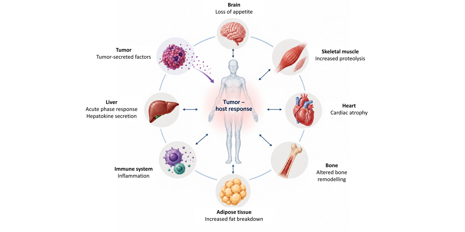
Cancer cachexia is driven by tumor- and host-derived signals that disrupt whole-body homeostasis and induce coordinated changes across multiple organs and tissues, including skeletal muscle, adipose tissue, heart, liver, brain and the immune system.
Understanding disease across the organism
Identifying these signals is only part of the picture. We also need approaches that capture how they reshape tissues across the body. Conventional analyses often focus on individual tissues or selected regions and can therefore miss coordinated responses occurring elsewhere in the body. We use tissue clearing and light-sheet microscopy together with computational image analysis to identify disease-associated changes in neural, immune and tissue architecture without preselecting specific regions.
Our previous work has shown the value of this organism-wide perspective. Applying whole-body analysis to obesity, we uncovered widespread remodeling of peripheral nerves (Figure 2) and immune cells across multiple tissues. This included structural alterations in facial sensory nerves that were associated with impaired sensory function. By combining these spatially resolved structural findings with spatial proteomics, we were able to link altered nerve architecture to local molecular changes and identify pathways associated with nerve remodeling. These studies demonstrate how organism-wide imaging can reveal unexpected sites of disease-associated alterations and connect structural phenotypes to their underlying molecular programs.
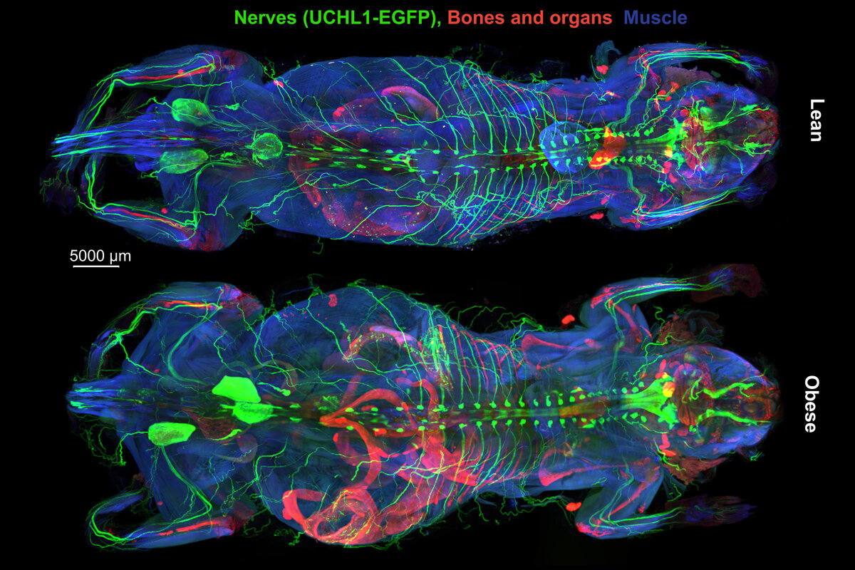
3D reconstructions of vDISCO-cleared lean and obese mice imaged by light-sheet fluorescence microscopy, showing Uchl1-eGFP+ peripheral nerves (Kaltenecker et al., Nature, 2026).
In our lab, we will build on this approach to understand how disease-associated signals are translated into coordinated responses across the organism. By combining organism-wide phenotyping with molecular and functional analyses, we aim to identify which tissues and cell types respond to disease-associated signals, uncover the mechanisms driving their remodeling and establish their contribution to disease.
More broadly, we are interested in the principles by which local molecular programs give rise to systemic disease states, and whether these principles are shared across different metabolic disorders.
Selected Publications
- Kaltenecker D*, Horvath I*, Al-Maskari R*, Chen Y*, Kolabas Z*, Hoeher L, Todorov M, Minde D, Kapoor S, Turhan S, Kuemmerle L, Steinke H, Wohlgemuth T, Ali M, Kofler F, Morigny P, Geppert J, Jeridi D, Wittmann B, Luo J, Shit S, Cigankova C, Kolenic V, Gür N, Aydeniz E, Yücecan A, Ertürk M, Simons L, Pan C, Piraud M, Rueckert D, Rohm M, Hellal F, Elsner M, Bhatia H, Bechmann I, Menze B, Herzig S, Paetzold J, Berriel Diaz M, Ertürk A. A deep-learning framework reveals whole-body perturbations at cell level. Nature 655, 1016–1026 (2026). DOI: 10.1038/s41586-026-10535-2.
- Kaltenecker D*, Fisker Schmidt S*, Weber P, Loft A, Morigny P, Machado J, Geppert J, Saul KB, Benedikt P, Molocea CE, Scott R, Haase K, Martignoni ME, Alfaro AJ, Chow KK, Simoes E, Pinhata Otoch J, Lima JDCC, Swanton C, Spielmann N, Hrabé de Angelis M, Elsner M, Ertürk A, Dyar KA, Rohm M, Prokopchuk O, Jamal-Hanjani M, Seelaender M, Backs J, Herzig S#, Berriel Diaz M#. Functional liver genomics identifies hepatokines promoting wasting in cancer cachexia. Cell 188, 4549–4566.e22 (2025). DOI: 10.1016/j.cell.2025.06.039.
- Kaltenecker D*, Al-Maskari R*, Negwer M*, Hoeher L, Kofler F, Zhao S, Todorov M, Rong Z, Paetzold JC, Wiestler B, Piraud M, Rueckert D, Geppert J, Morigny P, Rohm M, Menze BH, Herzig S, Berriel Diaz M#, Ertürk A#. Virtual reality-empowered deep-learning analysis of brain cells. Nature Methods 21, 1306–1315 (2024). DOI: 10.1038/s41592-024-02245-2.
- Morigny P, Vondrackova M, Ji H, Brejchova K, Krakovkova M, Makris K, Trubacova R, Samanci TF, Kaltenecker D, Ng SP, Karthikaisamy V, Chrysostomou SE, Bidovec A, Ponce-de-Leon M, Krauss T, Seeliger C, Prokopchuk O, Martignoni ME, Claussnitzer M, Hauner H, Schweiger M, Bindels LB, Berriel Diaz M, Herzig S, Lutter D, Kuda O#, Rohm M#. Multi-omics profiling of cachexia-targeted tissues reveals a spatio-temporally coordinated response to cancer. Nature Metabolism 8, 237–259 (2026). DOI: 10.1038/s42255-025-01434-3. #Co-corresponding authors.
- Machado J, Karthikaisamy V, Mohr H, Kaltenecker D, Benedikt P, Morigny P, Mhamane A, Geppert J, Fumo AR, Haase K, Simoes E, Lima JDCC, Georgiadi A, Krüger A, Pinhata Otoch J, Martignoni ME, Baracos VE, Jamal-Hanjani M, Seelaender MCL, Prokopchuk O, Szendrödi J, Rohm M, Herzig S, Berriel Diaz M. Tumor-secreted ADAMTSL4 activates latent TGFβ1 to drive cancer cachexia. Cancer Discovery (2026), online ahead of print. DOI: 10.1158/2159-8290.CD-26-0045.
Join us
- Master students and Post-docs: Contact Doris Kaltenecker with a letter of intent detailing why you want to join the lab.
- PhD students: Calls open 1 March and 1 September, apply here;
Vienna BioCenter PhD Program